Posterior fossa is a tight infratentorial compartment which harbors vital centers like cerebellum, pons and medulla. Anesthesia for Posterior Fossa Surgery is employed in carious types of space-occupying lesions, i.e., cerebellar tumours, cerebellopontine angle lesions, medulloblastoma, brainstem glioma occur in this area. The patients with posterior fossa tumor presents with features of raised intracranial pressure, brain stem dysfunction, cardiorespiratory abnormalities and preoperative/postoperative lower cranial nerve involvement. Besides this, cerebrovascular lesions like posterior inferior cerebellar artery aneurysm and basilar top aneurysm can also occur in the posterior fossa.
 Due to difficulty in approaching highly vascular posterior fossa aneurysms, these aneurysms are currently being managed non-surgically by the interventional neuroradiologist. Guglilemi detachable coils (GDC) are deployed into the aneurysmal sac subsequently leading to embolization of the aneurysm and prevention of rebleeding. However, patients with space-occupying lesions need to be operated early to prevent permanent damage to the vital structures.
Perioperative considerations for Anesthesia for Posterior Fossa Surgery are as follows:
- These tumours are approached using lateral, park-bench, prone or sitting position. Each surgical approach has its own advantages and disadvantages.
- Preoperative work up should include echocardiography to rule out patent foramen ovale. It has been found that 20—25% of the normal persons may have patent foramen ovale. The risk of paradoxical air embolism is high in these patients especially if the surgery is being performed in the sitting position. Similarly, elderly patients planned for surgery in the prone or sitting should be advised X-ray cervical spine. Presence of degenerative cervical spine diseases increases the risk of cervical cord injury with subsequent paraplegia in this group of patients.
- Small children with craniopharyngioma during Anesthesia for Posterior Fossa Surgery are usually operated in prone position. Use of armored endotracheal tube minimizes the risk of intraoperative tube kinking. Patient should be carefully positioned prone to facilitate adequate mechanical ventilation. Undue abdominal pressure should be avoided with the use of lateral rolls, because it can lead to venocaval compression. Pressure points should be properly padded. Patients with preoperative facial nerve involvement are unable to close eyes properly. Meticulous perioperative eye care (eye lubricants, adhesive tapes, eye pads) is required to prevent keratitis and loss of vision.
- In adults, surgery used to be done in sitting position because it provided excellent venous and cerebrospinal fluid drainage. However, the current status of this position is controversial because of the risk of cardiovascular instability and VAE.
Incidence of Doppler-detectable VAE during craniotomies in adults as well as children ranges from 20— 40%. Hence, appropriate monitoring should be available for early detection of VAE. Monitoring of somatosensory and motor evoked potentials before and after making the surgical position which can affect the cervical cord minimizes the risk of positioning complications. Once VAE is detected prompt management should be done as discussed in the previous section. Other reported perioperative complications of the sitting position include midcervical quadriplegia, systemic hypotension and macroglossia.
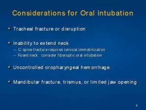
- Patients with foramen magnum tumours and ArnoldChiari malformations may have limited neck extension making airway management difficult. Previous history of skull base surgery and temporalis muscle flap reconstruction can also lead to difficulty in intubation because of the associated pseudoankylosis of the mandible. Other factors associated with the difficulty in maintaining a patent airway are: pre-existing cranial nerve deficits especially bulbar palsy, flexed intraoperative sitting position, overzealous nasal packing, surgical insult to brainstem, perioral oedema and/or rapidly expanding intracranial haematoma.
- Labile hypertension in the pre-surgical period may lead to rightward shift of the cerebral autoregulatory curve, hence higher arterial pressures ( > 20—30% of baseline) will be required to maintain adequate perfusion of brain parenchyma.
- Surgery demands brain retraction or non eloquent brain resection to prevent postoperative herniation. However, prolonged retraction can affect venous outflow leading to brain tissue damage and haemorrhagic infarctions. Excessive retraction of posterior temporal lobe can lead to tearing of vein of Labb’e and severe haemorrhagic temporal lobe oedema. Adequate bone removal, CSF drainage and use of diuretics can aid in adequate exposure without excessive brain retraction.
- At the end of surgery, patient should be thoroughly evaluated. Cerebral air embolism, hypoxic-ischaemic injury or prolonged surgical manipulation can lead to focal neurological deficits, stroke and coma. These patients may require ventilatory support in the postoperative period.